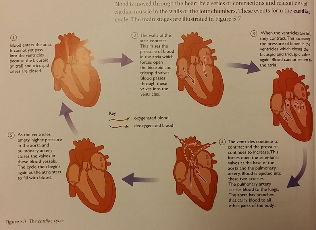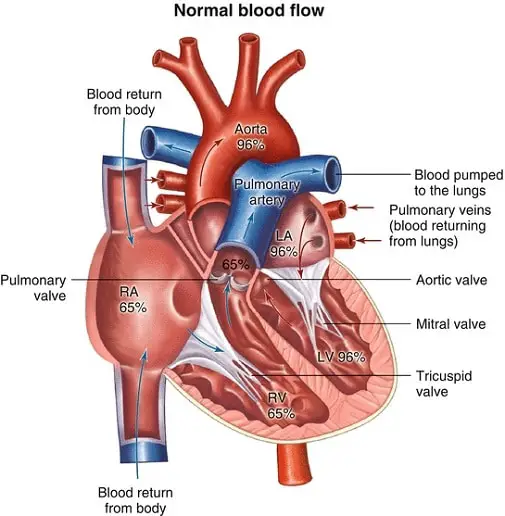

Once blood is pumped out of the left ventricle and into the aorta, the aortic semilunar valve (or aortic valve) closes preventing blood from flowing backward into the left ventricle. This blood passes through the bicuspid valve or mitral valve (the atrioventricular valve on the left side of the heart) to the left ventricle where the blood is pumped out through aorta, the major artery of the body, taking oxygenated blood to the organs and muscles of the body. The left atrium then receives the oxygen-rich blood from the lungs via the pulmonary veins.


After it is filled, the right ventricle pumps the blood through the pulmonary arteries, by-passing the semilunar valve (or pulmonic valve) to the lungs for re-oxygenation.Īfter blood passes through the pulmonary arteries, the right semilunar valves close preventing the blood from flowing backwards into the right ventricle.
/human-heart-circulatory-system-598167278-5c48d4d2c9e77c0001a577d4.jpg)
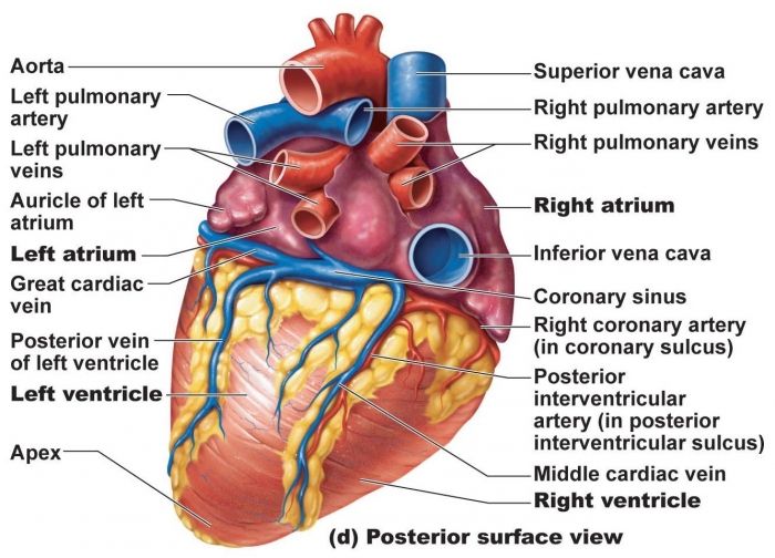
The valve separating the chambers on the left side of the heart valve is called the bicuspid or mitral valve. This deoxygenated blood then passes to the right ventricle through the atrioventricular valve or the tricuspid valve, a flap of connective tissue that opens in only one direction to prevent the backflow of blood. In addition, the right atrium receives blood from the coronary sinus which drains deoxygenated blood from the heart itself. The right atrium receives deoxygenated blood from the superior vena cava, which drains blood from the jugular vein that comes from the brain and from the veins that come from the arms, as well as from the inferior vena cava which drains blood from the veins that come from the lower organs and the legs. The atria are the chambers that receive blood, and the ventricles are the chambers that pump blood. There is one atrium and one ventricle on the right side and one atrium and one ventricle on the left side. In humans, the heart is about the size of a clenched fist, and it is divided into four chambers: two atria and two ventricles. One-way valves separate the four chambers. The heart is primarily made of a thick muscle layer, called the myocardium, surrounded by membranes. Since the right side of the heart sends blood to the pulmonary circuit, it is smaller than the left side which must send blood out to the whole body in the systemic circuit, as shown in Figure 1. The heart muscle is asymmetrical due to the distance blood must travel in the pulmonary and systemic circuits.


 0 kommentar(er)
